Just a quick one today. Nala didn’t seem to interested in trotting along beside me. She kept stopping and looking at me, saying “What are we doing out here? Let’s go eat!”. It was her regular breakfast time after all. I think I’ll try again later this afternoon.
This past weekend, I strapped on the running shoes again. It’s been a long time since I stopped running. 9 years ago when I moved to Detroit was when I stopped. There simply weren’t any safe runnable areas around me. Then I moved to Charleston SC, where again there weren’t a lot of decent bikable or runnable areas that didn’t have traffic speeding by at 40 or 50 mph. Sure, there are a lot of runners around. But by then, it had been a few years, and work was busy, so I never got started back up. Since then I’ve packed on about 30 extra pounds and have been missing my biking and running since then. There’s also this thing about the weather too. I’m not sure how people do it around here, running around in 40°+ weather. I can hardly walk out the door to the car in the driveway without having all the life sucked out of me by the heat and humidity!
Now we have Nala, and I decided it was finally time to get off my lazy fat ass and start running again, both as a way to help burn off some of that excess lab puppy energy, and for me to finally get back into shape and drop some of this extra blubber.
Runners and coaches tell people that keeping a running log is a good way of keeping yourself motivated and tracking your progress. That’s what this space is for, my running log.
A long, long time ago during my undergrad days, one of the requirements for my 4th year physics lab course (way back in 1990 or so) was to do some kind of research project. The project my lab partner and I settled on was to investigate the use of copper filters in the x-ray beam to reduce skin entrance exposure to pediatric cardiac cath patients. For the past few years it’s something that’s been pretty standard in fluoroscopic equipment now (although not something I can claim any credit for, I don’t think). I thought for posterity, I’d post our original write-up here.
Disclaimer: This work is from a project I did for one of my undergraduate labs a few years ago. The only review process this project has undergone was being scrutinized by my supervisor for this project and by the TA who marked it. Check my bibliography for other published papers on the subject.
If you’d like to comment on or criticize this, email me at eugenemah@gmail.com (or just leave a comment)
Abstract
Pediatric patients undergoing cardiac catheterization procedures can be subjected to large skin entrance exposures from both fluoroscopy and cine modes. To reduce the radiation exposure to the patient while minimizing loss in image quality, a small amount of copper filtration is added. In conjunction with standard radiation protection procedures (shielding and minimizing procedure time) the reduction in patient exposure can be reduced quite significantly. Preliminary phantom investigations demonstrate a decrease in exposure of up to 70% with the addition of 0.2 cm of copper filtration and little image quality degradation.
Introduction
Cardiac catheterization procedures involving pediatric patients can subject both the patient and operators to high radiation exposures. Because of the small size of the patients involved, children receive higher gonadal and thyroid exposures from scattered radiation. Children are also more sensitive to the effects of radiation than adults.
Several methods of reducing patient dose have been suggested such as minimizing procedure time, reducing the x-ray field size or placing lead shielding underneath the patient. Newer pulsed fluoroscopy systems where the x-ray beam is pulsed at the same rate as the video refresh rate of the monitor (30-60 pulses/second) has also shown to result in a significant saving in patient dose as well as improving image quality [7, 8, 13].
In keeping with the principle of ALARA, a method of reducing patient exposure by introducing additional beam filtration is proposed and investigated in this work. The additional filtration reduces radiation exposure by hardening the beam and removing the low energy x-rays which do not contribute to the formation of the x-ray image. Since the average kVp for pediatric patients is between 50-60 kVp, this should result in a large reduction in patient exposure without significant degradation in image quality. Aluminum filtration was also examined to compare the reduction in exposure between copper and aluminum. A similar method employed by den Boer et al [8] demonstrated a 55% reduction in patient exposure and 69% reduction in operator exposure with the combination of a high-output pulsed fluoroscopy and 0.4 mm of copper filtration.
Materials and Methods
Skin entrance exposures in typical cardiac catheterization procedures were examined for a Siemens cardiac catheterization laboratory equipped with a Siemens Polydoros 100 high frequency generator, Bicor biplane C-arms with variable FOV image intensifiers, Cinematic AEC 35 mm cine camera and Hicor digital imaging system.
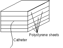
Figure 1: Phantom used for skin entrance exposure phantom
A simple phantom consisting of a number of Lucite sheets was assembled to mimic a small patient approximately 6 cm thick. A small length of catheter was placed in the center of the phantom to serve as a reference marker and a rough indication of the image quality as the filters were added.
Skin entrance exposures for both fluoroscopy and cine were measured with a Keithly electrometer and a small ion chamber placed underneath the phantom. For fluoroscopy and cine, the skin entrance exposures for various amounts of aluminum and copper filtration were measured and graphed. Aluminum thicknesses ranging from 0-4.5 cm and copper thicknesses from 0-2.0 mm were used.
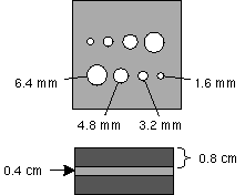
Figure 2: Low contrast phantom
The low contrast phantom (Figure 2) was made up of a 0.4 cm thick plate of aluminum with 8 holes of 4 different sizes, 1.6, 3.2, 4.8 and 6.4 mm in diameter. This plate was sandwiched between two 0.8 cm blocks of aluminum so that the holes in the aluminum sheet were 2% of the total thickness (2.0 cm).
Figure 3: High contrast phantom
The high contrast resolution phantom is a 0.5 cm thick block of plastic with 8 metal grids of varying line pair densities embedded in it. Line pair densities range from 20 lpi (lines/inch) to 60 lpi.
Both phantoms were placed on the table top 30 cm away from the image intensifier and 40 cm above the x-ray tube and examined using fluoroscopy and cine. Image quality was evaluated using each phantom for a variety of filter thicknesses using the fluoroscopy display monitors and cine film.
Results
Image Quality
For the low contrast phantom and up to 0.2 mm of copper filtration added, all 4 holes were still easily visible. With 0.4 mm of copper filtration, the smallest hole (1.6 mm) became barely visible and was no longer visible with 0.5 mm of copper filtration.
High contrast resolution (HCR) was measured at about 28 lpi (lines/inch) with up to 0.2 mm of copper filtration. At 0.4 mm and 0.5 mm of copper filtration, HCR dropped to 24 lpi.
Exposure Rates
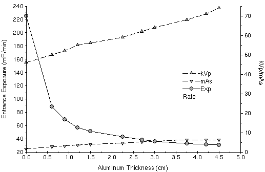
With the phantom placed on the table and the ion chamber underneath the phantom, fluoroscopic skin entrance exposure rates in mR/min were measured first with no aluminum filtration, and then with increasing amounts of aluminum added to the x-ray tube. X-ray techniques (kVp and mAs) were also recorded. Table 1 shows the results with the results illustrated graphically in Figure 4.
Table 1: Fluoroscopic skin entrance exposure rates for Al filter
Amt of Al (cm)
kVp
mAs
Exposure rate (mR/min)
0
46
1.6
225
0.6
50
2.7
89
0.9
52
3.1
69.2
1.2
55
3.6
57
1.5
56
4
51.3
2.25
59
4.6
43
2.7
62
5.2
38.6
3
64
5.6
36.2
3.75
68
6.3
32.8
4.2
71
6.3
31.5
4.5
74
6.3
30.8
Fluoroscopic skin entrance exposure rates for Al filter
Figure 4: Fluoro skin entrance w/ Al graph
Figure 4 illustrates the dramatic decrease in skin entrance exposure with the addition of just 0.6 cm of additional aluminum filtration. The additional filtration results in a 60% reduction in patient exposure and only a slight increase in x-ray technique (from 46 kVp to 50 kVp and 1.6 mAs to 2.7 mAs).
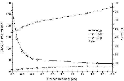
The same measurements were performed using copper filtration, with the results tabulated in Table 2 and illustrated in Figure 5.
Table 2: Fluoroscopic skin entrance exposure rates for Cu filter
Amt of Cu (mm)
kVp
mAs
Exposure rate (mR/min)
0
46
1.8
267
0.1
48
2.3
141
0.2
52
3
88
0.3
54
3.4
70
0.4
56
3.9
57
0.5
58
4.3
53
1.5
58
6.3
38.4
1.7
71
6.4
35.3
2
75
6.4
34.7
Fluoroscopic skin entrance exposure rates for Cu filter
Figure 5: Fluoro skin entrance w/ Cu graph
The reduction in patient exposure is nearly 50% with 0.1 mm of copper and 67% with 0.2 mm with very little change in x-ray technique (Figure 5). For larger filter thicknesses, the reduction in patient exposure is small relative to the increase in x-ray technique and the expected degradation in image quality resulting from the higher techniques.
Table 3 shows the cine skin entrance exposure with copper filtration, illustrated in Figure 6.
Table 3: Cine skin entrance exposure rates for Cu filter
Amt of Cu (mm)
kVp
mAs
Exposure rate (R/min)
0
50.5
28
3.32
0.2
54
29
0.64
0.4
60.5
31
0.4
Cine skin entrance exposure rates for Cu filter
Figure 6: Cine skin entrance w/ Cu graph
For cine procedures where exposure rates can be in the R/min range, 0.2 mm of copper filtration resulted in an 80% decrease in patient exposure with little change in technique. Unfortunately, due to time limitations and the arrival of a patient, fluoroscopy exposure rates were not obtained for copper filter thicknesses between 0.5 mm and 1.5 mm and only 3 measurements could be made in cine mode.
Conclusion
Inserting a small amount of additional filtration is a simple and effective way of obtaining significant exposure savings for pediatric patients undergoing cardiac catheterization procedures where they may be subjected to high radiation doses. From a practical viewpoint, copper would appear to be the most ideal filter, since copper provides much greater reduction in patient exposure and is much thinner than the equivalent amount of aluminum. An additional 0.2 mm of copper filtration appears to provide an optimum amount of filtration which maximizes the radiation exposure reduction and minimizes the degradation in image quality.
Bibliography
Rueter FG, “Physician and patient exposure during cardiac catheterization”, Circulation, 58:134-139 1978
Martin EC, Olson A, “Radiation exposure to the paediatric patient from cardiac catheterization and angiocardiography”, Br J Radiol, 53:100-106 1979
Wu JR, Huang TY, Wu DK, Hsu PC, Weng PS, “Radiation exposure of pediatric patients and physicians during cardiac catheterization and balloon pulmonary valvuloplasty”, Am J Cardiol, 68:221-225 1991
Martin EC, Olson AP, Seeg CN, Casarella WJ, “Radiation exposure to the pediatric patient during cardiac catheterization and angiocardiography”, Circulation, 64:153-157 1981
Waldman JD, Rummerfield PS, Gilpin EA, Kirkpatrick SE, “Radiation exposure to the child during cardiac catheterization”, Circulation, 64:158-163 1981
Schueler BA, Julsrud PR, Gray JE, Stears JG, Wu KY, “Radiation exposure and efficacy of exposure reduction techniques during cardiac catheterization in children”, Am J Roentgenol, 162:173-177 1994
den Boer A, de Feyter PJ, Hummel WA, Keane D, Roelandt JRTC, “Reduction of radiation exposure while maintaiing high quality fluoroscopic images during interventional cardiology using novel x-ray tube technology with extra beam filtering”, Circulation, 89:2710-2714 1994
Li LB, Kai M, Takano K, Ikeda S, Matsuura M, Kusama T, “Occupational exposure in pediatric cardiac catheterization”, Health Phys, 69:261-264 1995
Johnson LW, Moore RJ, Balter S, “Review of radiation safety in the cardiac catheterization laboratory”, Cathet Cardiovasc Diagn, 25:186-194 1992
Miller SW, Castronovo Jr FP, “Radiation exposure and protection in cardiac catheterization laboratories”, Am J Cardiol, 55:171-176 1985
Faulkner K, Love HG, Sweeney JK, Bardsley RA, “Radiation doses and somatic risk to patients during cardiac radiological procedures”, Br J Radiol, 59:359-363 1986
Holmes DR, Wondrow MA, Gray JE, Vetter RJ, Fellows JL, Julsrud PR, “Effect of pulsed progressive fluoroscopy on reduction of radiation dose in the cardiac catheterization laboratory”, J Am Coll Cardiol, 15:159-162 1990
Wilma is starting to finish it’s pummeling of the Yucutan and heading off to Florida. Fortunately for those in Florida, NHC isn’t forecasting Wilma to get much stronger and to pass through pretty rapidly.
From today’s 5AM NHC discussion for Wilma:
AIR FORCE RECON OBSERVATIONS INDICATE THAT THE INNER CORE OF WILMA…WHICH WAS DISRUPTED DUE TO ITS INTERACTION WITH LAND…IS BECOMING SLIGHTLY BETTER DEFINED. HOWEVER THE EYE IS STILL QUITE LARGE…ABOUT 65 N MI ACROSS…AND RAGGED. THE CENTRAL PRESSURE HAS NOT CHANGED MUCH SINCE THE HURRICANE EMERGED FROM NORTHEAST YUCATAN…AND THE CURRENT INTENSITY OF 85 KT IS BASED ON PEAK FLIGHT-LEVEL WINDS OF 91 KT TO THE SOUTH OF THE CENTER…AND SOME RESPECT FOR A CENTRAL PRESSURE OF 961 MB. SATELLITE IMAGERY SHOWS THAT THE CLOUD PATTERN IS ELONGATED ALONG A NNE-SSW AXIS WITH MOST OF THE DEEP CONVECTION DISPLACED TO THE NORTH-NORTHEAST OF THE CENTER. THIS MAY BE A HARBINGER OF INCREASING SHEAR…BUT IT IS ASSUMED THAT WILMA’S INNER CORE WILL BE ABLE TO AT LEAST TEMPORARILY REDEVELOP…AND THAT SOME RESTRENGTHENING WILL OCCUR TODAY. HOWEVER…THE GLOBAL MODELS INDICATE A SUBSTANTIAL INCREASE IN VERTICAL SHEAR WILL OCCUR AS WILMA NEARS THE FLORIDA PENINSULA… WHICH…ALONG WITH POSSIBLE ENTRAINMENT OF DRIER LOWER-LEVEL AIR… WOULD LEAD TO WEAKENING. SINCE THE TROUGH INTERACTING WITH WILMA IS QUITE STRONG…THE CYCLONE IS FORECAST TO BECOME EXTRATROPICAL AT A RELATIVELY LOW LATITUDE BY ABOUT 48 HOURS.
Yesterday, TS Alpha (2005) blossomed in the middle of the Carribean and will be dumping a bunch of rain over the Dominican Republic and Haiti today. After that, it looks like it will be carried out into the Atlantic along with Wilma without much additional strengthening.
So apparently my undead friend Oreo has communicated with the land of the living through one of my friends to say that yes, he really is getting married this time. No idea when or where. Communicating with the dead is never very reliable after all. And then there’s the matter of wallets.


